Effect of stress on brain inflammation and multiple sclerosis
this is the re-uploaded version of my previous Naver Blog Posting at 2022-05-21
Effect of stress on brain inflammation and multiple sclerosis
Cite: Karagkouni, A., Alevizos, M., & Theoharides, T. C. (2013). Effect of stress on brain inflammation and multiple sclerosis. Autoimmunity reviews , 12(10), 947-953. Link
Abstract
- MS: multiple sclerosis
- demyelinating, autoimmune disease ← inflammation of CNS
- stress→MS: ???
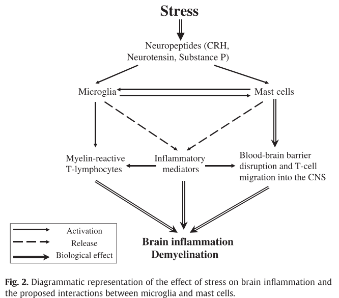
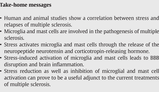
- propose that neuropeptides secreted under stress: activate microglia&mast cells
- CRH, NT(neurotnsin)
- ⇒maturation/activation of T17 autoimmune cells, disruption of BBB, T cell entry into the CNS
- ⇒promote brain inflammation, contributing to MS pathology
- novel therapeutic approach: reduction of stress & inhibition by flavonoids
Introduction
MS: autoimmune disease ← brain inflammation, demyelination, axonal loss
trigerring factors: infections, toxins, immunizations, trauma, sunlight, hormone…
stress can worsen immunity/brain inflammation → important in the pathogenesis of MS, neuropsychiatric disorders in general
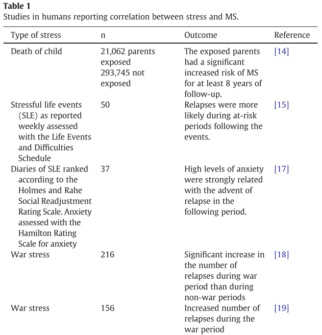
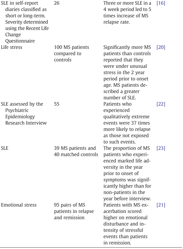
Correlation between stress and MS
Human studies
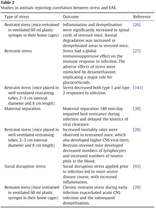
Animal studies
mouse model: TMEV(Theiler’s murine encephalomyelitis virus) infection
induce EAE: experimental allergic encephalomyelitis
Involvement of microglia and mast cells

microglia play an important role in the pathogenesis of MS
MRP14, 27E10 expressed(acute stage inflammatory macrophage markers) ⇒ intense microglia infiltration in acute MS cases
hypoxia-like lesion existing MS subtype: microglial activation is prominent, precedes T-cell infiltration & demyelination
death by MS → extensive oligodendrocyte apoptosis & microglial activation in the relative absence of T-cells
Roles of Microglia
act as antigen-presenting cells for naive T-cells
expanding the number of encephalitogenic Th1 cells
produce glutamate, NO → direct effect on the death of neurons
NO: cytotoxic effect on the endothelium → contribute to BBB disruption
dying oligodendroglial cells: recruit microglia
→ induce contact-dependent oligodendroglial death in the presence of IFN-γ activation
rich source of ROS & various pro-inflammatory cytokineschemokines/proteases
- Role of Microglia in the termination of the inflammatory reaction
-
suppress lymphocyte reactivity through NO release
strong accumulation of CD 163(+) microglia with anti-inflammatory effects was found in acute active MS lesions and at the rim of chronic active lesions
possibly involved in the resolution of the inflammation
phagocytose apoptotic T-cells(seems to be defective in MS)
in mice with EAE
microglial activation persists in chronic phase
T cell infiltrates are predominant during the acute phase of the disease
microglia participate in the pathogenesis of EAE: phagocytosing myelin + releasing TNF-α, IL-1, IL-6, chemokines
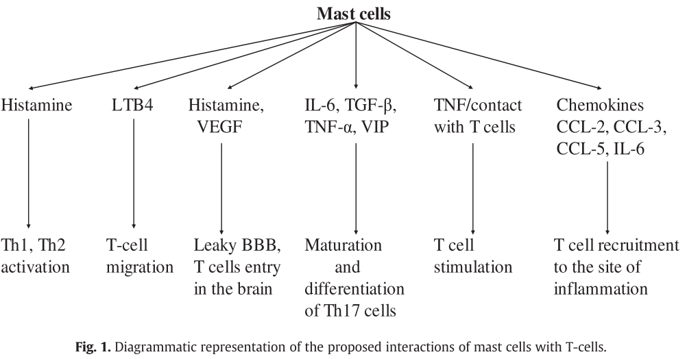
Th1 and Th2
Th1 cells, macrophages: brain infiltration
Th2 cells, mast cells: associated with allergic reactions
MS plaque at mast cell reported, could stimulate demyelination directly
Possible mechanisms to translate stress in MS risk
- actue stress permissive effect on MS exacerbation by facilitating BBB breakdown
- chronic stress lead to glucocorticoid resistance → immunce cells less responsive to regulatory control by cortisol
Stress and the HPA axis
Stress → CRH hypothalamic secretion → activate HPA(hypothalamic-pituitary-adrenal) axis
→glucocorticoids releasion → suppress immune response
In this context…
may be due to dysfunctional HPA axis because of reduced production of adrenal steroids
patients data is consistent… (absence of a normal cortisol response)
also, glococorticoid resistance development due to stress exposure can be a cause
Stress, microglia and mast cells
CRH and NT
- CRH의 pro-inflammatory effects affects brain microvessels directly → activate mast cells → BBB permeability increase
- CRH and NT which are secreted under stress synergistically stimulate mast cells → vascular permeability increase, BBB disruption
- NT stimulates mast cell secretion of VEGF(vascular endothelial GF) →increase BBB permeability
- NT induce expression of CRHR-1(CRH Receptor-1)
Stress activates microglia
- in vivo experiment evidence
- CRH: induce proliferation and TNF-α release in microglia
- microglia express NTR3 → MIP-2, MIP-1, IL-1β, TNF-α
- SP receptors detected: activation lead to the activatio of NF-κB TF
- human - microglia also produce SP → activate mast cells
- microglia respond to pro-inflammatory signals released from mast cells
- mast cell tryptase: induces microglial activation and pro-inflammatory mediator release(TNF-α, IL-6, ROS)
Conclusion
treatment?
- stress-management program
- diazepam, alprazolam
-
CRH antagonist
- microglia, mast cell as therapeutic targets? ← no clinically available inhibitors
- natural flavonoids(quercetin, luteolin, apigenin): anti-oxidant, anti-inflammatory effects, suppress TNF-α, IL-6 expression and release from microglia, mast cell activation, release of cytokines
- luteolin, quercetin: decrease myelin phagocytosed by macrophages, reduce EAE
- luteolin: inhibits mast cell-dependent T cell activation
- apigenin: sensitizes activated human T cells to apoptosis, inhibits auto-antigen-presenting cells necessary for the expansion and activation of Th17 cells in lupus
- propolis(flavonoid-containing): inhibits IL-6, TGF-β induced TH17 differentiation in vitro
- luteolin: inhibit activated peripheral blood mononuclear cells and synergistic effect with IFN-β
- luteolin may be reasonable!!
Leave a comment